There is a correlation of tobacco-product use and various conditions of the teeth, the jawbone, and the soft tissue of the mouth. The appearance, site, and incidence of these conditions can vary considerably with which type of tobacco product is used. Although smoking is the commonest mode of tobacco use in the United States, the popularity of tobacco chewing and snuff dipping is on the rise.
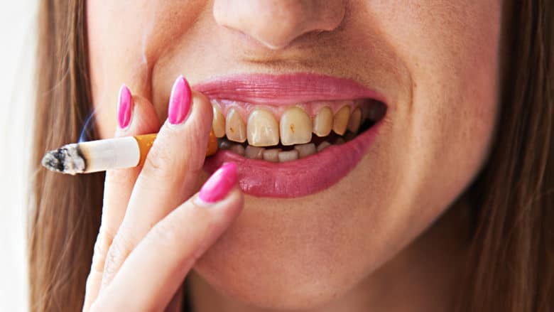
Smokers tend to have discolorations, ranging from dark brown to black, at the gumline from tar and other byproducts of tobacco combustion. Among pipe smokers, pipe-stem erosion of teeth is common and can result in an open bite—in short, a gap between upper and lower front teeth. Acute necrotizing ulcerative gingivitis (ANUG)—a progressive disease of the gum characterized by bleeding, swelling, and pain of that tissue—occurs mainly among young heavy smokers who neglect oral hygiene. How ANUG originates has not been fully described, but (a) ischemia due to nicotine-induced vasoconstriction and (b) a tar-accumulation-induced excess of the buildup of dental plaque may be responsible for the disease.
There is a correlation not only of smoking and an elevated risk of developing periodontal disease, but also of smoking and extra bone loss in persons with periodontal disease. By affecting adversely the action of neutrophils, smoking impairs immune reactions and, therefore, recovery. Thus, routine gum surgery for reducing perio pockets is less effective in smokers than in nonsmokers.
Smokers’ lips and the linings of their cheeks are commonly sites both of burns and of white patches, usually either flat or slightly raised, with red striations. The patches disappear with cessation of the habit.
Heavy smokers commonly develop a condition called “black hairy tongue,” which is characterized by discoloration and elongation of the papillae of the tongue. This condition calls for a biopsy, and correcting it may require surgical and/or other therapy.
Pipe smokers commonly develop nicotinic stomatitis—a condition characterized by red or grayish-white discoloration of the palate (the roof of the mouth) with numerous red dots. (Dots of this kind result from inflammation of salivary ducts in the palate.) Such stomatitis is generally not treated as precancerous, and cessation of the habit usually results in its quick resolution.
Heavy smokers sometimes develop painful palatal sores due to hot gases. Such sores should be tested by biopsy for cancer. A biopsy is critical in cases of another smoking-related lesion, the leukoplakia (the word is basically a combination of two words that together denote “white flat area”). Three to six percent of leukoplakias become cancers.
Tobacco use is the principal cause of malignant tumors of the mouth. Smoking a packful of cigarettes daily or routinely using chewing tobacco quadruples one’s risk of developing such a tumor. Oral cancer and pharyngeal (throat) cancer constitute a major cause of cancer-related death in the U.S., and cases of these cancers constitute about 3-4 percent of American cancer cases. The mortality of cancer of the mouth and/or pharynx exceeds that of cervical cancer or melanoma.
Of oral and pharyngeal cancers in the U.S., 96 percent are carcinomas and four percent are sarcomas. Approximately 90 percent of the cases of oral cancer in the U.S. are cases of squamous cell carcinoma. Oral carcinomas most commonly occur at the back of either narrow side of the tongue; they also commonly occur on the floor of the mouth. Therefore, the dentist must not only feel the underside of the tongue and the floor of the mouth; he or she must also hold the tongue outside the mouth to facilitate visual examination. Oral carcinomas typically have a red or white appearance, and those that are not ulcerated almost never cause physical pain. Speech alteration, persistent hoarseness, or a chronic cough is sufficient reason to suspect metastasis.
Carcinomas of the lip, which usually appear as painless ulcers, constitute 25-30 percent of oral cancers. A biopsy should be done on any lip ulcer that has lasted for at least two weeks. Verrucous (wartlike) carcinomas constitute 4.5-9 percent of oral carcinomas. They appear as gray or white protuberances, grow slowly, and occur mostly among male smokers older than 65 years.
Treatment of oral cancer of any kind usually involves both surgery and irradiation but not chemotherapy.
There are two basic forms of smokeless tobacco: snuff and chewing tobacco. Dry snuff is inhaled through the nose, while moist snuff is placed inside the mouth. Chewing tobacco is coarser than snuff and comes as loose contents of pouches and small packages, as small blocks, and as rope-like twists of dried tobacco leaves. Because chewing tobacco can abrade enamel and is high in sugar (sugar is responsible for 30-40 percent of its weight), dental cavities can result from its use, especially along the roots of those teeth adjacent to which the wad of tobacco is placed. Tooth decay at tobacco-wad sites can be major.
Smokeless tobacco is widely recognized among physicians and medical scientists as a carcinogen. Verrucous carcinomas and squamous cell carcinomas are the cancers that its users most commonly develop.
Historically, dentists have used scalpels to take samples of cancerous-looking abnormalities. But there is an inexpensive and easier mode of biopsy, called a “brush biopsy.” In this procedure, which requires neither anesthesia nor suture, the dentist removes some cells from the abnormality with a tiny brush whose bristles are stiff. These cells are placed on a slide, which is sent to facilities where such cells are tested by a computer. Slides with suspicious cells are examined by a pathologist. If the pathologist decides that the cells the dentist submitted are abnormal, such information is faxed to the dentist within a few days, whereupon the dentist refers the patient to a specialist.
In recent studies, all cancers were detected, without any false negatives, through the procedure described above.
The oral health consequences of routine use of tobacco products of any kind are serious. Everyone should demand a thorough oral cancer screening at least once a year during a regular check-up.
